蛋白酪氨酸磷酸酶CD148抗体
产品名称: 蛋白酪氨酸磷酸酶CD148抗体
英文名称: DEP1
产品编号: 2567
产品价格: null
产品产地: 上海
品牌商标: 雅吉
更新时间: null
使用范围: WB ELISA IHC-P
上海雅吉生物科技有限公司
- 联系人 :
- 地址 : 上海市闵行区元江路5500号第1幢5658室
- 邮编 :
- 所在区域 : 上海
- 电话 : 158****3937 点击查看
- 传真 : 点击查看
- 邮箱 : yajikit@163.com
| 中文名称 | 蛋白酪氨酸磷酸酶CD148抗体 |
| 别 名 | CD 148; CD148; CD148 antigen; Density enhanced phosphatase 1; Density-enhanced phosphatase 1; DEP 1; DEP-1; HPTP eta; HPTPeta; Human density enhanced phosphatase 1; Protein tyrosine phosphatase eta; Protein tyrosine phosphatase receptor type J; Protein tyrosine phosphatase receptor type J polypeptide; Protein-tyrosine phosphatase eta; Protein-tyrosine phosphatase receptor type J; PTPJ; Ptprj; PTPRJ_HUMAN; R PTP Eta; R-PTP-eta; R-PTP-J; Receptor type tyrosine protein phosphatase eta; Receptor-type tyrosine-protein phosphatase eta; SCC 1; SCC1. |
| 研究领域 | 细胞生物 信号转导 激酶和磷酸酶 |
| 抗体来源 | Rabbit |
| 克隆类型 | Polyclonal |
| 交叉反应 | Human, Mouse, (predicted: Rat, Rabbit, ) |
| 产品应用 | WB=1:500-2000 ELISA=1:500-1000 IHC-P=1:100-500 (石蜡切片需做抗原修复) not yet tested in other applications. optimal dilutions/concentrations should be determined by the end user. |
| 分 子 量 | 147kDa |
| 细胞定位 | 细胞膜 |
| 性 状 | Liquid |
| 浓 度 | 1mg/ml |
| 免 疫 原 | KLH conjugated synthetic peptide derived from human PTPRJ:865-970/1337 |
| 亚 型 | IgG |
| 纯化方法 | affinity purified by Protein A |
| 储 存 液 | 0.01M TBS(pH7.4) with 1% BSA, 0.03% Proclin300 and 50% Glycerol. |
| 保存条件 | Shipped at 4℃. Store at -20 °C for one year. Avoid repeated freeze/thaw cycles. |
| PubMed | PubMed |
| 产品介绍 | The protein encoded by this gene is a member of the protein tyrosine phosphatase (PTP) family. PTPs are known to be signaling molecules that regulate a variety of cellular processes, including cell growth, differentiation, mitotic cycle, and oncogenic transformation. This PTP possesses an extracellular region containing five fibronectin type III repeats, a single transmembrane region, and a single intracytoplasmic catalytic domain, and thus represents a receptor-type PTP. This protein is present in all hematopoietic lineages, and was shown to negatively regulate T cell receptor signaling possibly through interfering with the phosphorylation of Phospholipase C Gamma 1 and Linker for Activation of T Cells. This protein can also dephosphorylate the PDGF beta receptor, and may be involved in UV-induced signal transduction. Multiple transcript variants encoding different isoforms have been found for this gene. [provided by RefSeq]. Function: Tyrosine phosphatase which dephosphorylates or contributes to the dephosphorylation of CTNND1, FLT3, PDGFRB, MET, RET (variant MEN2A), KDR, LYN, SRC, MAPK1, MAPK3, EGFR, TJP1, OCLN, PIK3R1 and PIK3R2. Plays a role in cell adhesion, migration, proliferation and differentiation. Involved in vascular development. Regulator of macrophage adhesion and spreading. Positively affects cell-matrix adhesion. Positive regulator of platelet activation and thrombosis. Negative regulator of cell proliferation. Negative regulator of PDGF-stimulated cell migration; through dephosphorylation of PDGFR. Positive regulator of endothelial cell survival, as well as of VEGF-induced SRC and AKT activation; through KDR dephosphorylation. Negative regulator of EGFR signaling pathway; through EGFR dephosphorylation. Enhances the barrier function of epithelial junctions during reassembly. Negatively regulates T-cell receptor (TCR) signaling. Upon T-cell TCR activation, it is up-regulated and excluded from the immunological synapses, while upon T-cell-antigen presenting cells (APC) disengagement, it is no longer excluded and can dephosphorylate PLCG1 and LAT to down-regulate prolongation of signaling. Subunit: Monomer. Interacts with CTNNB1 (phosphorylated) and JUP (phosphorylated). Interacts with FLT3 (phosphorylated). Interacts with GAB1 and GRB2. Subcellular Location: Cell membrane. Tissue Specificity: Expressed in the promyelocytic cell line HL60, the granulocyte-macrophage colony-stimulating factor-dependent leukemic cell line F-36P, and the interleukin-3 and erythropoietin-dependent leukemic cell line F-36E. Expressed predominantly in epithelial cells and lymphocytes. Enhanced expression at high cell density. Post-translational modifications: N- and O-glycosylated. Similarity: Belongs to the protein-tyrosine phosphatase family. Receptor class 3 subfamily. Contains 9 fibronectin type-III domains. Contains 1 tyrosine-protein phosphatase domain. SWISS: Q12913 Gene ID: 5795 Database links: Entrez Gene: 5795 Human Omim: 600925 Human SwissProt: Q12913 Human Unigene: 318547 Human Important Note: This product as supplied is intended for research use only, not for use in human, therapeutic or diagnostic applications. |
| 产品图片 | 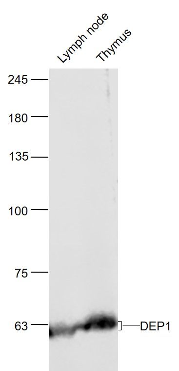 Sample: Sample:Lymph node (Mouse) Lysate at 40 ug Thymus (Mouse) Lysate at 40 ug Primary: Anti-DEP1 (bs-2567R) at 1/1000 dilution Secondary: IRDye800CW Goat Anti-Rabbit IgG at 1/20000 dilution Predicted band size: 60'100 kD Observed band size: 60 kD 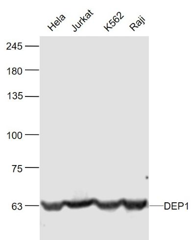 Sample: Sample:Hela(Human) Cell Lysate at 30 ug Jurkat(Human) Cell Lysate at 30 ug K562(Human) Cell Lysate at 30 ug Raji(Human) Cell Lysate at 30 ug Primary: Anti-DEP1 (bs-2567R) at 1/1000 dilution Secondary: IRDye800CW Goat Anti-Rabbit IgG at 1/20000 dilution Predicted band size: 60'100 kD Observed band size: 60 kD 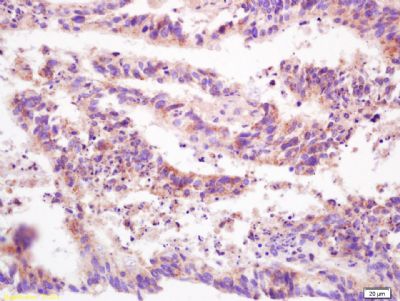 Tissue/cell: human colon carcinoma; 4% Paraformaldehyde-fixed and paraffin-embedded; Tissue/cell: human colon carcinoma; 4% Paraformaldehyde-fixed and paraffin-embedded;Antigen retrieval: citrate buffer ( 0.01M, pH 6.0 ), Boiling bathing for 15min; Block endogenous peroxidase by 3% Hydrogen peroxide for 30min; Blocking buffer (normal goat serum,C-0005) at 37℃ for 20 min; Incubation: Anti-PTP/PTPase/CD148 Polyclonal Antibody, Unconjugated(bs-2567R) 1:200, overnight at 4°C, followed by conjugation to the secondary antibody(SP-0023) and DAB(C-0010) staining 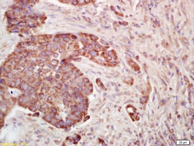 Tissue/cell: human lung carcinoma; 4% Paraformaldehyde-fixed and paraffin-embedded; Tissue/cell: human lung carcinoma; 4% Paraformaldehyde-fixed and paraffin-embedded;Antigen retrieval: citrate buffer ( 0.01M, pH 6.0 ), Boiling bathing for 15min; Block endogenous peroxidase by 3% Hydrogen peroxide for 30min; Blocking buffer (normal goat serum,C-0005) at 37℃ for 20 min; Incubation: Anti-PTP/PTPase/CD148 Polyclonal Antibody, Unconjugated(bs-2567R) 1:200, overnight at 4°C, followed by conjugation to the secondary antibody(SP-0023) and DAB(C-0010) staining |