弹性蛋白抗体
产品名称: 弹性蛋白抗体
英文名称: Elastin
产品编号: 1756
产品价格: null
产品产地: 上海
品牌商标: 雅吉
更新时间: null
使用范围: WB ELISA IHC-P IHC-F Flow-Cyt IF
上海雅吉生物科技有限公司
- 联系人 :
- 地址 : 上海市闵行区元江路5500号第1幢5658室
- 邮编 :
- 所在区域 : 上海
- 电话 : 158****3937 点击查看
- 传真 : 点击查看
- 邮箱 : yajikit@163.com
| 中文名称 | 弹性蛋白抗体 |
| 别 名 | alpha Elastin/Tropoelastin; Elastin isoform a; ELN; ELN_HUMAN; elastin isoform m precursor; FLJ38671; FLJ43523; Supravalvular aortic stenosis; Tropoelastin; Williams Beuren syndrome; Williams syndrome region; ADCL1; SVAS; WBS; WS. |
| 研究领域 | 肿瘤 免疫学 神经生物学 转录调节因子 |
| 抗体来源 | Rabbit |
| 克隆类型 | Polyclonal |
| 交叉反应 | Human, Mouse, Rat, |
| 产品应用 | WB=1:500-2000 ELISA=1:500-1000 IHC-P=1:100-500 IHC-F=1:100-500 Flow-Cyt=1μg /test IF=1:100-500 (石蜡切片需做抗原修复) not yet tested in other applications. optimal dilutions/concentrations should be determined by the end user. |
| 分 子 量 | 70kDa |
| 细胞定位 | 细胞外基质 分泌型蛋白 |
| 性 状 | Liquid |
| 浓 度 | 1mg/ml |
| 免 疫 原 | KLH conjugated synthetic peptide derived from human Elastin:681-786/786 |
| 亚 型 | IgG |
| 纯化方法 | affinity purified by Protein A |
| 储 存 液 | 0.01M TBS(pH7.4) with 1% BSA, 0.03% Proclin300 and 50% Glycerol. |
| 保存条件 | Shipped at 4℃. Store at -20 °C for one year. Avoid repeated freeze/thaw cycles. |
| PubMed | PubMed |
| 产品介绍 | This gene encodes a protein that is one of the two components of elastic fibers. Elastic fibers comprise part of the extracellular matrix and confer elasticity to organs and tissues including the heart, skin, lungs, ligaments, and blood vessels. The encoded protein is rich in hydrophobic amino acids such as glycine and proline, which form mobile hydrophobic regions bounded by crosslinks between lysine residues. Degradation products of the encoded protein, known as elastin-derived peptides or elastokines, bind the elastin receptor complex and other receptors and stimulate migration and proliferation of monocytes and skin fibroblasts. Elastokines can also contribute to cancer progression. Deletions and mutations in this gene are associated with supravalvular aortic stenosis (SVAS) and autosomal dominant cutis laxa. [provided by RefSeq, Aug 2017]. Function: Major structural protein of tissues such as aorta and nuchal ligament, which must expand rapidly and recover completely. Molecular determinant of the late arterial morphogenesis, stabilizing arterial structure by regulating proliferation and organization of vascular smooth muscle. Subunit: The polymeric elastin chains are cross-linked together into an extensible 3D network. Forms a ternary complex with BGN and MFAP2. Interacts with MFAP2 via divalent cations (calcium > magnesium > manganese) in a dose-dependent and saturating manner. Subcellular Location: Secreted, extracellular space, extracellular matrix. Note=Extracellular matrix of elastic fibers. Tissue Specificity: Expressed within the outer myometrial smooth muscle and throughout the arteriolar tree of uterus (at protein level). Also expressed in the large arteries, lung and skin. DISEASE: Defects in ELN are the cause of cutis laxa, autosomal dominant, type 1 (ADCL1) . A connective tissue disorder characterized by loose, hyperextensible skin with decreased resilience and elasticity leading to a premature aged appearance. Face, hands, feet, joints, and torso may be differentially affected. Additional variable clinical features are gastrointestinal diverticula, hernia, and genital prolapse. Rare manifestations are pulmonary artery stenosis, aortic aneurysm, bronchiectasis, and emphysema. Similarity: Belongs to the elastin family. SWISS: P15502 Gene ID: 2006 Database links: Entrez Gene: 2006 Human Entrez Gene: 13717 Mouse Omim: 130160 Human SwissProt: P15502 Human SwissProt: P54320 Mouse Unigene: 647061 Human Important Note: This product as supplied is intended for research use only, not for use in human, therapeutic or diagnostic applications. |
| 产品图片 | 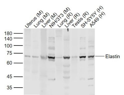 Sample: Sample:Lane 1: Uterus (Mouse) Lysate at 40 ug Lane 2: Lung (Mouse) Lysate at 40 ug Lane 3: Liver (Mouse) Lysate at 40 ug Lane 4: NIH/3T3 (Mouse) Cell Lysate at 30 ug Lane 5: Lung (Rat) Lysate at 40 ug Lane 6: Liver (Rat) Lysate at 40 ug Lane 7: Testis (Rat) Lysate at 40 ug Lane 8: SH-SY5Y (Human) Cell Lysate at 30 ug Lane 9: A549 (Human) Cell Lysate at 30 ug Primary: Anti-Elastin (bs-1756R) at 1/1000 dilution Secondary: IRDye800CW Goat Anti-Rabbit IgG at 1/20000 dilution Predicted band size: 66 kD Observed band size: 68 kD 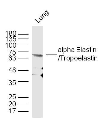 Sample: Lung (Mouse) Lysate at 30 ug Sample: Lung (Mouse) Lysate at 30 ugPrimary: Anti- alpha Elastin/Tropoelastin (bs-1756R) at 1/300 dilution Secondary: IRDye800CW Goat Anti-Mouse IgG at 1/10000 dilution Predicted band size: 70 kD Observed band size: 68 kD 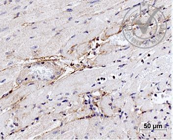 Images provided the Independent Validation Program (badge number 28751): Formalin-fixed and paraffin embedded mouse heart labeled with Rabbit Anti-alpha Elastin Polyclonal Antibody (bs-1756R) at 1:1000 4℃ temperature overnight 4℃followed by conjugation to secondary antibody. Images provided the Independent Validation Program (badge number 28751): Formalin-fixed and paraffin embedded mouse heart labeled with Rabbit Anti-alpha Elastin Polyclonal Antibody (bs-1756R) at 1:1000 4℃ temperature overnight 4℃followed by conjugation to secondary antibody.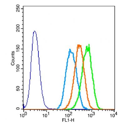 The figure annotation: The blue histogram is unstained cells.The green histogram is cells stained with Rabbit Anti-alpha Elastin/Tropoelastin antibody (bs-1756R)plus secondary antibody. The figure annotation: The blue histogram is unstained cells.The green histogram is cells stained with Rabbit Anti-alpha Elastin/Tropoelastin antibody (bs-1756R)plus secondary antibody.Controls: Positive control: A549 cells .Isotype control: Cell lines treated with rabbit IgG (bs-0295P)instead of the primary antibody to confirm that primary antibody binding is specific.Secondary only control: Both cell lines treated with Goat Anti-rabbit IgG/FITC antibody (bs-0295G-FITC) to confirm no background signal produced from secondary antibody alone. 1ug in 100uL 1 X PBS containing 0.5% BSA. |